Equipment
The following equipment for is available for small animal imaging (mice, rats, guinea pigs, rabbits and other similar sized animals).
nanoScan PET/CT Small Animal Imager
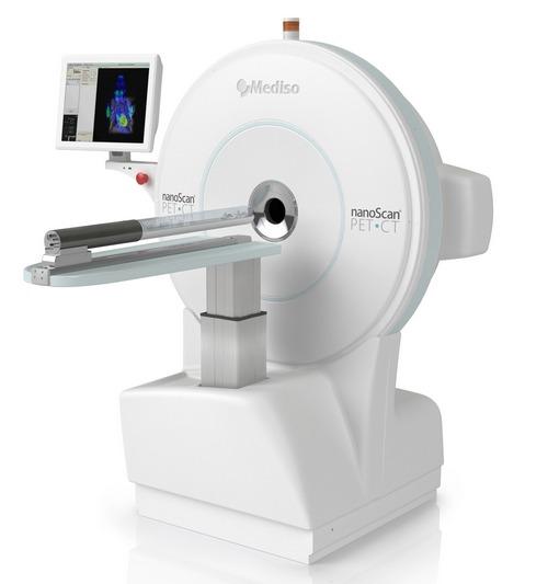
Advanced Positron Emission Tomography (PET) with integrated CT system dedicated for preclinical imaging. Unmatched resolution and sensitivity for small and medium size animals (mice to monkey heads) with 16 cms gantry bore diameter and 12 cms transaxial field of view (FOV) for both modalities. High-density, ultra-small LYSO detectors in tight packing provide the highest resolution PET subsystem (≤ 0.7 mm resolution) in the industry, uniform over the entire FOV, and quantitative accuracy better than 97%. Increased crystal thickness for high sensitivity (7%). The CT offers a large detector surface area, with helical scanning (30 cms scanning length), variable zoom (up to 7.6x magnification), and a high power X-ray source (80W), which can perform a whole-body mouse scan in ~4 minutes, with real time reconstruction with 100 micron voxel size.
This scanner is assisted use only and there is a user fee of $400/hour. These fees are refundable up to 48 hours prior to the scheduled time if it needs to be cancelled. User fee includes anesthesia supplies, use of the animal bed and environmental controls, remote data retrieval and use of advanced image analysis on our workstations. However, it does not include the cost of any radiotracers or other material (if required) for imaging
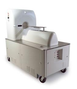
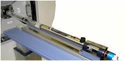
NanoSPECT/CT Small Animal Imager (CT currently not available)

Dual-Detector System has 2 broadband NaI(Tl) SPECT detectors with transaxial FOV of up to 20 cm and axial field-of-view of up to 27 cm due to helical scan data acquisition. User interchangeable multi-pinhole aperture plates and parallel hole collimators allow multiplexed, multi-pinhole high resolution (<0.5 mm), high sensitivity (>250-400 cps/MBq) SPECT imaging. Collimators and aperture plate sets for high-sensitivity or high-resolution imaging are also available. Detector hardware and software includes multi-channel analysis of SPECT emissions with energies ranging from 25 keV to 250 keV (I-125 to In-111) and simultaneous acquisition of dual-isotope images in separate energy windows with multi-channel analysis for spectral unfolding.
This scanner is assisted use only and there is a user fee of $175/hour per hour for each scheduled hour for the SPECT. These fees are refundable up to 48 hours prior to the scheduled time if it needs to be cancelled. User fee includes anesthesia supplies, use of the animal bed and environmental controls, remote data retrieval and use of advanced image analysis on our workstations. However, it does not include the cost of any radiotracers or other material (if required) for imaging.
Philips Mosaic HP Small Animal PET Imager
CereTom 8-slice CT Imager
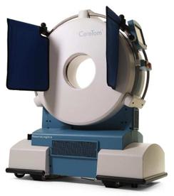
CereTom 8-slice clinical CT scanner with a large bore of 32.5 cm (318 mm), delivers the highest quality CT images and contrast perfusion scans. Due to its powerful X-ray source and rapid scan times, it can also perform angiography. The small animal scanning platform allows for imaging guinea pigs, rabbits and other small animals. The Barco’s Voxar 3D™ advanced visualization software package allows for 2D, 3D and MPR viewing. 1.25, 2.5, 5.0 and 10.0 mm slice thickness with CT data acquisition in multiple protocols is available. This imager complements the CT component of the NanoSPECT/CT but is primarily used for imaging large animals.
This scanner is assisted use only and there is a user fee of $175/hour for each scheduled hour*. These fees are refundable up to 48 hours prior to the scheduled time if it needs to be cancelled. User fee includes anesthesia supplies, use of the animal bed and environmental controls, remote data retrieval and use of advanced image analysis on our workstations. However, it does not include the cost of any contrast agents or other material (if required) for imaging.
Vevo 770 Small Animal Ultrasound System

The Vevo 770 system enables the visualization and quantification of small animal anatomical targets, hemodynamics and therapeutic interventions with resolution as low as 30 microns (high frequency scanhead) and frame rates up to 240 fps.
- B-mode (2D) imaging for anatomical visualization and quantification.
- M-mode for visualization and quantification of wall motion in cardiovascular research, single line acquisition allows for the very high-temporal (1000 fps) resolution necessary for analysis of LV function.
- Anatomical M-Mode for adjustable anatomical orientation in reconstructed M-Mode imaging.
- Pulsed-Wave Doppler (PW) for quantification of blood flow.
User fee is $135 per hour for each scheduled hour for both assisted and independent use. For independent use, the user must sign an agreement and undergo training prior to being on their own. Charging for independent use is $135 per hour beginning at the requested start time and ending when the scanner is turned off and/or the space is cleared if another researcher is waiting to use the ultrasound*. These fees are refundable up to 48 hours prior to the scheduled time if it needs to be cancelled. User fee includes anesthesia supplies, use of the animal bed and environmental controls, remote data retrieval and use of advanced image analysis on our workstations. However, it does not include the cost of any contrast agents or other material (if required) for imaging.
Ancillary Equipment and Services
- The animal bed and environmental controls are designed to provide monitoring and maintenance of ambient temperatures during imaging. They can also be used for containment of pathogen free animals.
- A high-speed workstation with ultra high speed graphics card and advanced image analysis software is available for reconstruction, data retrieval and advanced image analysis.
- All imaging data will be available in DICOM format. However, due to limited storage space, we highly encourage all users to copy their imaging data elsewhere. We do not provide guarantees for the integrity of these data for extended periods, after the completion of imaging.
The Center for Infection and Inflammation Imaging Research is located within the vivarium of the Cancer Research Building-II. To schedule a consultation or study, please call us at 410-614-3051 or email Sanjay Jain, MD and/or Mariah Klunk, BS. Also please visit our CrossLab site.